Alveokokkoz, or, as it is called, multilocular hydatid disease - a disease, which takes place in a person's severe chronic form. The primary focus is the liver injury. I.e, first the patient has liver alveococcosis, and only then - alveococcosis light. Symptoms of severe anthropozoonosis are not immediate, for a long time disease can occur in a latent form.
As a person gets infected
People get sick alveococcosis after accidental ingestion hexacanth (so called parasite eggs Alveococcus multilocularis). This can occur when taking untreated food, unwashed vegetables, fruit, green, and neglecting personal hygiene rules (washing hands before eating).
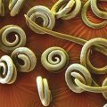
Parasitic origin of the disease first officially proved P. Virchow in 1856 city, proposing for this type of echinococcosis known as "multicam".

Alveococcosis can be attributed to the natural focal anthropozoonosis. Very poor regions in relation to the incidence of alveococcosis in the CIS are Kygrgyzstan, Kazakhstan and Uzbekistan, but, if it occurs in other areas, too, are not uncommon.
Etiology
For the full development cycle of the parasite has time to change the two hosts - from the intermediate host infection progresses to the final.
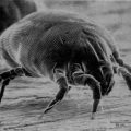
- Definitive host can become a fox, dogs, cherish, wolves and other predatory mammals, in the diet which includes small rodents. Tapeworm lives in the small section of the intestine, We are in a stage of mature individuals; in this period, the length is typically less than 4 cm.
- Then, during a 40-day period after the initial infection occurs parasite maturation terminal segments, They are filled with eggs and are released into the environment together with the excrement of the animal.
- hexacanth, falling in the environment in the water, on wild plants, wool of other animals, mushrooms - securely fastened and remain viable even in adverse conditions is critical, withstanding freezing to -21 ° C.
- small animals, swallow berries, mushrooms and seeds, affected helminth eggs, thereby to provide its larval stage of development, becoming the intermediate hosts of the parasite.
- If the affected berries, greenery, water, fruits and vegetables eats people, it also infects alveococcosis, becoming a biological dead end for parasite, which terminates at the larval stage of development of the helminth entire cycle. Person, Besides, can be infected with the violation to the rules of personal hygiene during butchering carcasses of animals or treatment of their skins.

- In the small intestine of human oncospheres is freed from the outer shell and their penetration into the mucosal capillaries.
- Then, with current venous they reach the target organ. Alveococcosis liver begins its development.
- The liver is the formation of larval helminths, which look like clusters of bubbles of small size, with the outside of the capsule and provided with chitin blastoderm within. Bubbles linked connective tissue between them, and can not by themselves actively replicate. Such alveococcus growth ensured by the presence in its capsule nucleation layer. Thanks to him, and made possible the appearance of new lesions and budding.
- multiply, bubbles are introduced into the parenchyma, forming infiltrates the surrounding tissue, and ensure the progression of exogenous development of helminth. There is a disseminated metastatic process.
Changes in the tissues
- fabrics, affected alveococcus, like tumor nodules, do not have clear boundaries and having a cellular structure in section. Each cell-alveolus has transverse dimensions to 5 mm and is filled with a gelatinous mass yellowish or brownish tint.
When conducting histology detected surrounded cuticular capsule type honeycomb structure. From within the elements of these structures lined with germinal shell.
Alveoli contain a capsule type brood, sometimes hooks, and scolexes. Reproduction alveococcus can be described as budding, an event such as the development of tumors with indomitable growth.
Capsules can invade nearby tissues. If the disease is at an early stage of existence, then during microscopy indicated parenchymal inflammation body to form a primary cell infiltration and reaction of granulomatous type.
In tissues, located close to the hearth, notes intense eosinophilia, plazmotsytoz, infiltration of lymphoid cells of the stroma, are manifestations of inflammatory response of vascular tissue - the arteries and veins.

- With regard to changes in the lungs, here metastatic alveococcosis outwardly resembles blastomatous tissue damage with a fairly dense areas, having an irregular shape and gray-white color. In the context, they do not differ from the liver, having the same type of cellular structure. Later in these areas develops coagulation necrosis.
When tissue destruction and disintegration of formed cavities, contain so-called detritus, gnoepodobnuyu curd type liquid. Adjacent tissues are hematoma, interalveolar partitions in some cases appear swollen.
With regard to the vascular tissue, here we see perivaskulity, endarteritis, necrosis and obliteration of vessel walls. Subsequently, there is a seal and sclerosis, and sometimes even calcification of the tissue.
Large cavity decay sometimes break, and their content falls into the nearby bronchi. Thus shatters almost all regions of the lung.
If there is a violation in the vessels of the circulatory and lymphatic systems, and entry to them alveococcus larvae, then the inevitable happens lymph node metastases, light, brain and bone marrow, as well as other organs and tissues.
Pathogenesis
- Peçenoçnıy alveokokkoz.
- Within months, and sometimes years disease, usually, developing an Email, without specific symptoms and remain undiagnosed. The parasite has the property to suppress the immune system of the patient. Allergy products helminth metabolism, arising in the affected organ, It does not cause human pain and intense inflammatory reactions.
- Manifestations of the disease is increasing with the growth of a parasitic formation in the target organ and adjacent organs.
- When the liver, spleen, lymph nodes and bone marrow are already heavy defeat, disease manifests as jaundice and symptoms of portal hypertension.

- Pulmonary alveococcosis develops:
BUT) as a consequence of the direct germination parasitic neoplasm of liver tissue through the diaphragmatic.
AT) Due hematogenous dissemination of parasitic scoleces through lower-portal system, heart and lung vessels. Such a mechanism of disease progression is seen in 1/4 alveococcosis ill patients.
- First, in the lung parenchyma changes occur infiltrative nature, resulting in varying degrees of severity focal pneumonia and bronchitis.
- When breaking through necrotic foci, arising in the central part of the large infiltrates, disease picture becomes similar to that for lung abscess.
- When distributing alveococcus on other systems of the body in the patient's condition deteriorates largely and acquires the picture, similar to the generalized blastomatozom.

Alveococcosis and diagnostics
More susceptible to other diseases of man and woman 20 to 50 years old. Occasionally the disease occurs in children and adults.
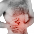
Complaints in the early stages of liver damage are reduced to a feeling of heaviness in the right upper quadrant and slightly pronounced pain in the epigastric.
Changes in the liver are often found by accident, or by examination, held due to the feeling of heaviness in the right hypochondrium. Sometimes a neoplasm increased density detected by palpation at the patients themselves.
- Primary disease declares itself a weakness, loss of appetite, epigastric pain of low intensity.
As the liver disease progresses jaundice and ascites is growing.
- When engaging in the pathological process diaphragm with subsequent infection of the lower segment of the right lung, there is respiratory discomfort and coughing with a minor amount of mucous or muco-purulent discharge.
- A break of metastatic lesions in the bronchi observed increase in body termperatury, cough with purulent sputum. Sometimes the patient has formed gall-bronchial fistula, sputum and acquires a characteristic color and texture.
At this stage, the disease symptoms are manifest not only the above. Along with them, continue to progress and hepatic manifestations of the disease.

- Generalized disseminated process, exciting the brain, kidneys and other organs, can change the picture of the disease is so, that complaints of sickness leave patients in the primary target organs by the wayside.
Diagnosis of the disease is difficult due to the lack of specific symptoms and the duration of its asymptomatic. In epidemicity areas recommended to periodically undergo immunological tests for the early detection and successful treatment of this debilitating disease.
- On physical patient study, an increase in liver size and change its density to a "rocky". Edge liver - smooth or has a fine-grained structure. Pain on palpation usually expressed slightly. This triad of symptoms is considered pathognomonic sign alveococcosis and is called the "Symptom Lyubimov" or "symptom of liver iron".

Sometimes patients have ikterichnost skin - but it is expressed softly, rather brown skin, than yellow, and has a grayish hue.
In some cases, as a symptom of portal hypertension, ascites in patients arises - the accumulation of fluid in the abdominal cavity.
but, physical examination data are not prognostically important, always requires additional laboratory tests.
The examination of the chest can be heard hard breathing, also it does not predict an important feature of this disease.
- The blood was:
- increased ESR,
- increased protein (gipyerprotyeinyemiya),
- gammaglobulinemia sustainable.
- More reliable results (order 90%) ELISA serology gives a positive reaction with, Phragmites, etc.. with hydatid diagnosticum.
- In the X-ray light alveococcosis manifested as various changes in the tissue, and combinations thereof: focal infiltrative, cavity, pneumonic and tumor. Characterized by calcification.
- CT and ultrasound - liver damage. Reminiscent of malignancy.
- Bronchoscopy - uninformative.
- Perytoneoskopyya, in some cases it is combined with laparotomy and taking a tissue sample to histology, It allows to differentiate from alveococcosis blastomatoza. It is associated with such technical difficulties, which suggests that it is not the most successful method of investigation and diagnosis of the disease.
Treatment
With involvement of the lung tissue, the disease is often poor prognosis, even if the therapy.
- If resection of the affected organ, for whatever reason, can not or no longer makes much sense, used chemotherapy, that inhibit the growth and spread of foci.
- In the nineties, 20 century have been made unsuccessfully attempts to use "tepal" drug, patients insertion / m in an amount 1 ml five times a day. Total held 5 courses at intervals of 7-14 days. The general condition of the patient thereby improving, because it reduces the activity alveococcus.
- Temporarily helps deter growth alveococcus, thereby extending the life of the patient, treatment with drugs mebendazole (following, how Vermoxum and telmoks), is carried out for a long period of repeated courses.
- If surgical treatment is carried out additional therapy 0,5 % formalin solution and 0,1 % rastvorami acres- and tripaflavina.
The operation often involves the removal of extensive liver portion with the capture portion of the diaphragm and parts of the right lung.
Forecast this disease is always careful. more, Unfortunately, adverse.